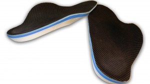
This first case is a patient who is a middle aged female who first started having pain in her right foot in 2002. She lived in another region of the country and first started having pain along the medial side of her foot (inside) which was diagnosed as a partial tear of the tibialis posterior tendon. This was treated with a cast immobilizer and an Arizona brace ( a custom large leather cast like brace that surrounds the foot and ankle and lower leg).
This was followed by a plastic arch device (UCBL) that was uncomfortable and did not resolve the problem. She was offered a tendon transplant to resolve the problem. She moved to New England.
Her pain persisted in spite of wearing braces. In 2003 she stopped wearing braces and was treated by a physical therapist for one year. The pain got worse and the foot collapsed. She saw another physician who recommended good orthotics which were prescribed.
Her diagnosis at this time was arthritis of the midfoot at the tarsal metatarsal joints as well as tibialis posterior tendon insufficiency (posterior tibial tendon dysfunction). Her foot at that point was painful all the time with considerable swelling and warmth in the midfoot and extreme difficulty walking.
At this time she presented to this office on referral of a physician friend. She was unable to walk, had a collapsed foot with considerable warmth and swelling and desperate to avoid surgery. Her x-rays were reviewed which showed conderable midfoot arthritis particularly in the first and second metatarsal cuneiform joint. The foot presented like a Charcot foot but in the absence of a neuropathy this was not the case. This woman was otherwise healthy.
On exam her calcaneal stance position (angle of heel to floor) was very high in valgus and her forefoot varus measured 17 degrees and her calf muscles were very short. All of these accounted for an extremely flat foot.
There semed to be some function of the posterior tibial tendon. Her options were to place her in a cast walker with and orthotic or to construct a short leg fiberglass bivalved and lined walking cast brace which I do at Childrens Hospital.

Custom Orthotic with high and thick lateral flanges, and comprehensive forefoot wedging.
Her foot however was quite swollen and I thought the best approach would be to construct a full length semi soft functional orthotic with a Brooks Ariel sneaker. We constructed the orthotics that same day and posted her to the full prescription of 7 degrees in the rearfoot and 17 degrees in the forefoot
under the 2nd thru 5th metatarsals. This in fact measured 24 degrees under the forefoot because we combine the rearfoot of 7 and the forefoot of 17. In order to accomplish this significant degree of posting it is necessary to construct a large lateral flange both in height and thickness.
The area under the first met head is also posted slightly less than under the second met. She was seen two weeks later with a significant decrease in swelling and pain. She was able to walk more comfortably. We continue to see her two months later and she continues to improve. She never needed a cast. Her strength has improved considerably in the tibialis posterior tendon. The arthritis in the first and second met cuneiform joints as burned out and she is no longer symptomatic there.
Comments are closed.